Myocardial mass and wall thickness
Feature summary
LV myocardial mass
Average ED/ES myocardial label volume converted to grams with the implemented myocardial density.
Hypertrophic cardiomyopathy (I42.2)Hypertension (I10)Disease badges are literature-context navigation only; not diagnoses, CardiacNexus classifiers, or validated phenotype-to-ICD associations.LV mass-to-volume ratio
Myocardial mass divided by LV end-diastolic volume as an LV concentricity marker.
Family: structuralUnit family: g/mLSource: Cine short-axis CMRPrimary output group: LV concentricityHypertensive heart disease (I11)Disease badges are literature-context navigation only; not diagnoses, CardiacNexus classifiers, or validated phenotype-to-ICD associations.AHA segment wall thickness
ED myocardial thickness for AHA segments 1-16 plus a global value.
Family: structuralUnit family: mmSource: Cine short-axis CMRPrimary output group: Segmental wall thicknessHypertrophic cardiomyopathy (I42.2)Amyloidosis (E85)Disease badges are literature-context navigation only; not diagnoses, CardiacNexus classifiers, or validated phenotype-to-ICD associations.Systolic wall thickening
Relative ED-to-ES increase in wall thickness for AHA segments and global myocardium.
Family: functionalUnit family: %Source: Cine short-axis CMRPrimary output group: Segmental thickeningAcute myocardial infarction (I21)Chronic ischemic heart disease (I25)Disease badges are literature-context navigation only; not diagnoses, CardiacNexus classifiers, or validated phenotype-to-ICD associations.
Myocardial mass and wall-thickness phenotypes describe LV myocardial amount, compact-myocardium thickness, and systolic thickening from cine short-axis segmentation.
- Modality
- Cine short-axis CMR
- UKB source
- Data Field 20209
- Pipeline step
- Myocardial segmentation, mass, wall thickness, thickening, and disparity extraction
- Outputs
- ventricular_volume.csv, wall_thickness.csv, myocardial contour landmarks, bullseye QC images
- Maturity
- Source-audited phenotype page
Clinical question
These outputs help distinguish chamber dilation from hypertrophy, concentric remodeling, regional thickening abnormalities, and trabeculation-like texture summaries. They are interpretation aids, not standalone HCM, amyloid, infarction, or hypertension classifiers.
Anatomical and physiological definition
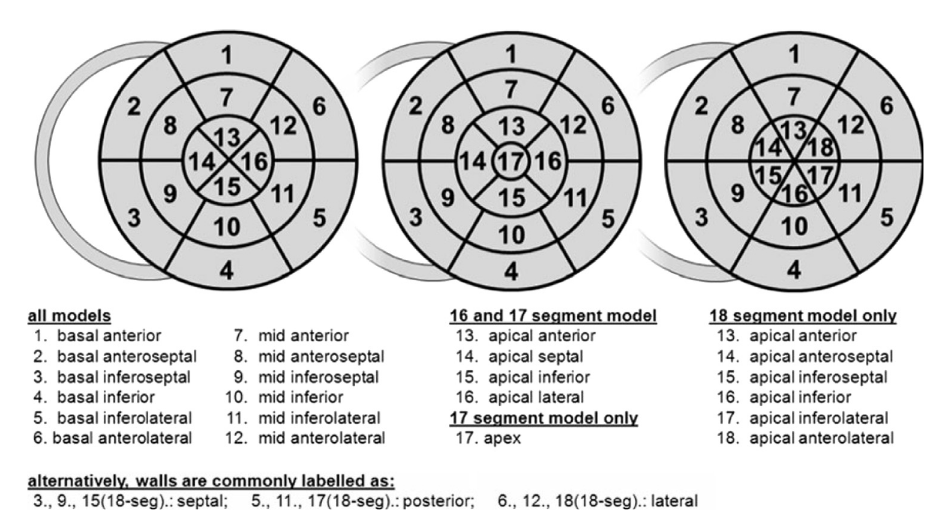
Myocardial mass is the amount of segmented LV myocardium converted from volume to grams. Wall thickness is measured between endocardial and epicardial contours. Systolic thickening is the relative increase from ED to ES. AHA segment labels 1-16 follow the conventional LV segment model; segment 17 is not emitted as a CSV column in the current wall-thickness output [1].
Source acquisition and UKB field
The source acquisition is UK Biobank cine short-axis CMR, Data Field 20209. Mass is emitted by the ventricular-volume script, while wall thickness, thickening, disparity, and fractal dimension are emitted by the wall-thickness script.
What exactly CardiacNexus measures
Myocardial mass
CardiacNexus averages ED and ES myocardial label volumes and multiplies by density 1.05 g/mL:
Copyable formula
Myo_Mass_g = ((Myo_EDV + Myo_ESV) / 2) * 1.05Mass-to-volume ratio
Copyable formula
LV_MV = Myo_Mass_g / LV_V_ED_mLWall thickness
The wall-thickness script computes ED myocardial thickness for AHA segments 1-16 plus a global summary using endocardial and epicardial contours from the short-axis segmentation.
| Variable | Definition | Output family |
|---|---|---|
wall_thickness_ED[0..15] | ED mean wall thickness for AHA segments 1-16 | Myo: Thickness (AHA_*) [mm] |
wall_thickness_ED[16] | global ED wall-thickness summary | Myo: Thickness (Global) [mm] |
wall_thickness_ES | ES wall-thickness vector used for thickening | internal intermediate plus ES QC bullseye |
thickening[0..15] | relative ED-to-ES segmental thickening | Myo: Thickening (AHA_*) [%] |
thickening[16] | global ED-to-ES thickening | Myo: Thickening (Global) [%] |
Systolic wall thickening
Copyable formula
Thickening = (WT_ES - WT_ED) / WT_ED * 100Disparity and fractal dimension
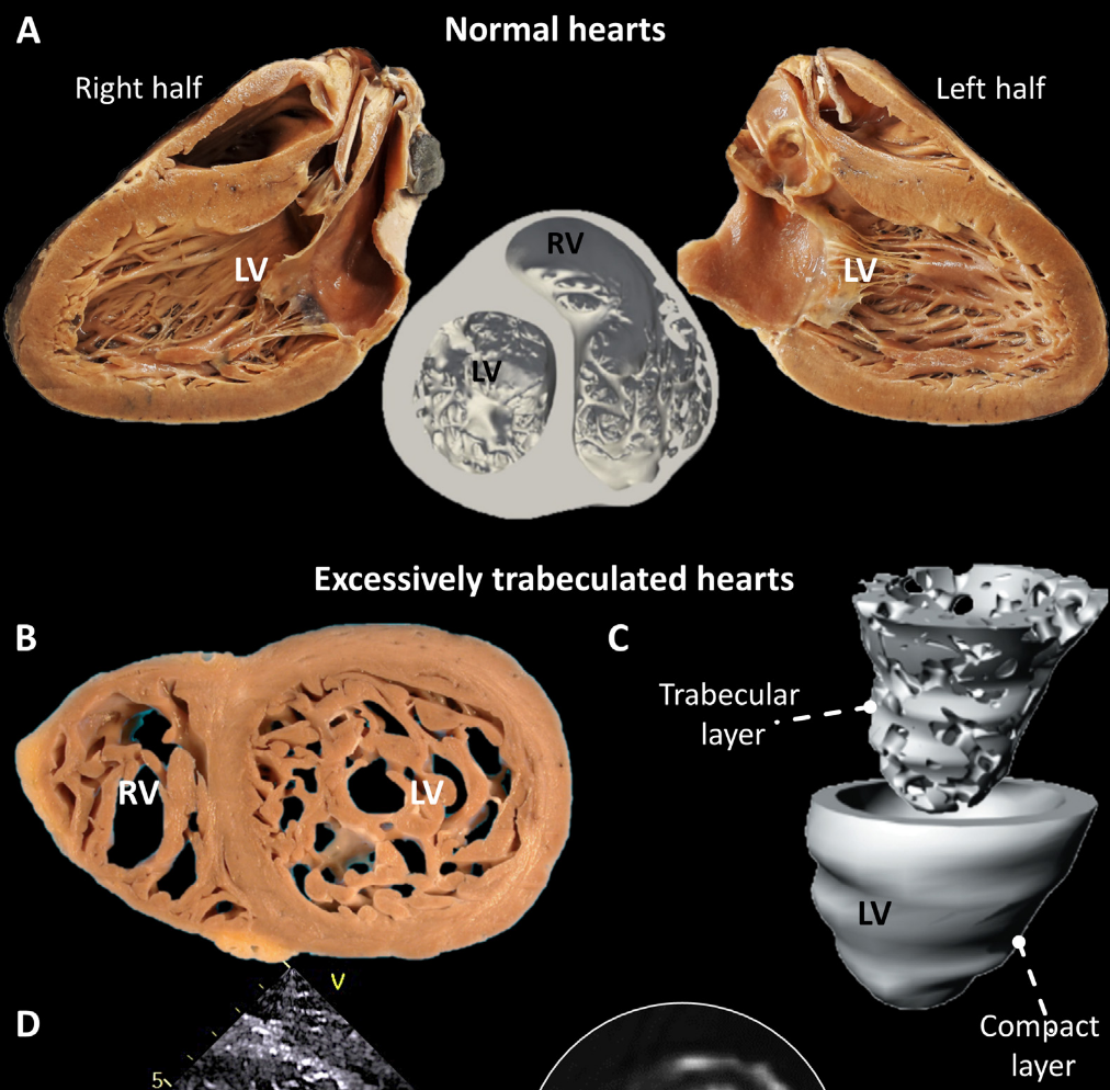
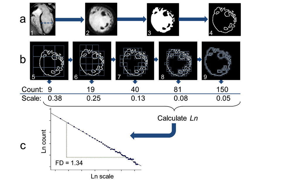
Radius and thickness motion disparity are computed from ED/ES endocardial and epicardial contours after BSA lookup and outlier guards. Fractal dimension is a global texture descriptor from the ED short-axis segmentation route; it is method-dependent and should not be treated as a trabeculation disease classifier.
Output columns and units
| Display family | Exact output column | Unit | Status | Schema note |
|---|---|---|---|---|
| Myocardial mass | Myo: Mass [g] | g | current | Emitted by ventricular_volume.csv |
| Indexed myocardial mass | Myo: Mass/BSA [g/m^2] | g/m² | current when BSA exists | BSA-dependent |
| LV concentricity | LV: M/V [g/mL] | g/mL | current | Mass divided by LV EDV |
| Global wall thickness | Myo: Thickness (Global) [mm] | mm | current | Emitted by wall_thickness.csv |
| Segment wall thickness | Myo: Thickness (AHA_1) [mm] through Myo: Thickness (AHA_16) [mm] | mm | current | AHA segment 17 is not emitted |
| Global thickening | Myo: Thickening (Global) [%] | % | current | ED-to-ES relative change |
| Segment thickening | Myo: Thickening (AHA_1) [%] through Myo: Thickening (AHA_16) [%] | % | current with outlier skip behavior | Values above the implemented threshold may be skipped |
| Motion disparity | Myo: Radius motion disparity | unitless | conditional current output | Requires BSA and passes outlier guard |
| Motion disparity | Myo: Thickness motion disparity | unitless | conditional current output | Requires BSA and passes outlier guard |
| Trabeculation texture | Myo: Fractal dimension | unitless | current | Global fractal dimension from ED segmentation route |
Segment output coverage
| Output family | Exact current columns |
|---|---|
| Segment wall thickness | Myo: Thickness (AHA_1) [mm], Myo: Thickness (AHA_2) [mm], Myo: Thickness (AHA_3) [mm], Myo: Thickness (AHA_4) [mm], Myo: Thickness (AHA_5) [mm], Myo: Thickness (AHA_6) [mm], Myo: Thickness (AHA_7) [mm], Myo: Thickness (AHA_8) [mm], Myo: Thickness (AHA_9) [mm], Myo: Thickness (AHA_10) [mm], Myo: Thickness (AHA_11) [mm], Myo: Thickness (AHA_12) [mm], Myo: Thickness (AHA_13) [mm], Myo: Thickness (AHA_14) [mm], Myo: Thickness (AHA_15) [mm], Myo: Thickness (AHA_16) [mm] |
| Segment systolic thickening | Myo: Thickening (AHA_1) [%], Myo: Thickening (AHA_2) [%], Myo: Thickening (AHA_3) [%], Myo: Thickening (AHA_4) [%], Myo: Thickening (AHA_5) [%], Myo: Thickening (AHA_6) [%], Myo: Thickening (AHA_7) [%], Myo: Thickening (AHA_8) [%], Myo: Thickening (AHA_9) [%], Myo: Thickening (AHA_10) [%], Myo: Thickening (AHA_11) [%], Myo: Thickening (AHA_12) [%], Myo: Thickening (AHA_13) [%], Myo: Thickening (AHA_14) [%], Myo: Thickening (AHA_15) [%], Myo: Thickening (AHA_16) [%] |
Output reconciliation
| Evidence layer | Result |
|---|---|
| Implementation source | 40 myocardial page-scope outputs documented here: 3 mass/concentricity rows, 17 wall-thickness rows, 17 thickening rows, 2 disparity rows, and 1 fractal-dimension row |
| Output inventory | ventricular_volume and wall_thickness inventories include the same 40 page-scope outputs |
| Phenotype dictionary | docs/data/phenotype_dictionary.yml links the same 40 outputs to this page |
| Page output table | all 40 output labels are listed above |
Required upstream inputs
sa.nii.gz,sa_ED.nii.gz,seg_sa.nii.gz,seg_sa_ED.nii.gz, andseg_sa_ES.nii.gz.- Short-axis segmentation QC passing
sa_pass_quality_control. - BSA lookup for indexed mass and disparity outputs.
Reference ranges with cohort and method context
| Feature | Source | Cohort | Reference value | Status | Note |
|---|---|---|---|---|---|
| LV mass | Petersen et al. UK Biobank CMR reference ranges [2] | UK Biobank CMR reference cohort | sex- and age-stratified source rows | Verified context source | Use segmentation convention context; not copied as CardiacNexus-specific thresholds |
| LV mass and wall thickness | Kawel-Boehm et al. CMR reference update [3] | multi-study CMR reference context | study-specific | Verified context source | Segment and papillary-muscle conventions vary |
| AHA segmentation | AHA segment nomenclature statement [1] | consensus nomenclature | not a normal range | Definition source | Used for segment naming, not normal limits |
| Fractal dimension | Captur et al. myocardial trabeculation fractal analysis [4] | MESA CMR context | method-dependent | Verified context source | Context only; current CardiacNexus global fractal-dimension route needs method-matched validation before thresholds |
Source-located registry status: reference_range_sources.yml maps LV mass and indexed-mass context to Petersen 2017 source tables, wall-thickness context to Kawel-Boehm 2020, and fractal-dimension context to Captur 2015. Numeric public thresholds are not promoted for wall thickness, thickening, or fractal dimension until CardiacNexus segmentation, papillary-muscle, AHA segment, and fractal-analysis conventions are matched row by row.
Disease interpretation
Higher LV mass and wall thickness are relevant to hypertrophic cardiomyopathy, hypertension, aortic stenosis, and infiltrative disease contexts [2] [3]. Reduced regional thickening is relevant to ischemic injury and cardiomyopathy contexts. Fractal dimension is trabeculation-method context [4]. These are literature-context badges only; CardiacNexus does not diagnose HCM, amyloid, infarction, or hypertension.
QC caveats and maturity boundary
Mass depends on myocardium label quality and basal/apical slice inclusion. Thickness and thickening depend on endocardial/epicardial contour stability, AHA segment assignment, and ED/ES segmentation consistency. Very high thickening values are skipped by implementation guards.
Implementation provenance
Mass and M/V are implemented in src/feature_extraction/Short_Axis_20209/eval_ventricular_volume.py. Wall thickness, thickening, radius/thickness disparity, and fractal dimension are implemented in src/feature_extraction/Short_Axis_20209/eval_wall_thickness.py.
| Feature family | Formula or computational route | Exact output columns | Source code file and function | Upstream dependencies | Conditional behavior | QC artifacts | Schema debt |
|---|---|---|---|---|---|---|---|
| Myocardial mass and index | average ED/ES myocardium label volume times density 1.05; indexed row divides by BSA | Myo: Mass [g], Myo: Mass/BSA [g/m^2] | eval_ventricular_volume.py; basic and indexed feature blocks | seg_sa.nii.gz, voxel spacing, BSA table | subject skipped if short-axis QC or BSA lookup fails before indexed outputs | seg_sa_ED.png, seg_sa_ES.png, ventricular volume QC | mass emitted from ventricular-volume artifact, not wall-thickness artifact |
| LV concentricity | myocardial mass divided by LV EDV | LV: M/V [g/mL] | eval_ventricular_volume.py; M/V block | valid LV ED volume and myocardial mass | inherits ventricular volume QC and BSA skip lifecycle | ventricular volume QC | row lives in ventricular_volume.csv |
| ED wall thickness | evaluate_wall_thickness(seg_sa_ED, nim_sa_ED) for AHA segments and global index | Myo: Thickness (Global) [mm], Myo: Thickness (AHA_1) [mm] through Myo: Thickness (AHA_16) [mm] | eval_wall_thickness.py; wall-thickness block; utils.cardiac_utils.evaluate_wall_thickness | sa_ED.nii.gz, seg_sa_ED.nii.gz, contour helpers | subject skipped if segmentation/QC inputs fail | thickness_ED.png, endocardial/epicardial VTK landmarks | AHA segment 17 is not emitted |
| Systolic thickening | (wall_thickness_ES - wall_thickness_ED) / wall_thickness_ED * 100 | Myo: Thickening (Global) [%], Myo: Thickening (AHA_1) [%] through Myo: Thickening (AHA_16) [%] | eval_wall_thickness.py; thickening block | seg_sa_ED.nii.gz, seg_sa_ES.nii.gz | segment rows skipped if value exceeds implemented high-thickening guard | thickness_ES.png, thickening.png | high values are omitted rather than clipped |
| Motion disparity | evaluate_radius_thickness_disparity on ED/ES contours with BSA | Myo: Radius motion disparity, Myo: Thickness motion disparity | eval_wall_thickness.py; disparity block | ED/ES contours and BSA lookup | rows skipped if BSA unavailable or outlier guard fails | contour landmarks and wall-thickness QC | method-dependent exploratory descriptor |
| Fractal dimension | slice-level fractal-dimension extraction summarized globally | Myo: Fractal dimension | eval_wall_thickness.py; fractal-dimension block; utils.cardiac_utils.fractal_dimension | ED segmentation route | current output if calculation succeeds | fractal-dimension visualization path | method-dependent trabeculation texture descriptor |
Source audit
- Formula and output claims were checked against the current ventricular-volume and wall-thickness scripts.
- Full page-scope output coverage was reconciled across source contracts, page text,
docs/data/output_column_inventory.yml, anddocs/data/phenotype_dictionary.yml. docs/data/reference_sources.ymlis present and used as the curated reference-source registry for this page.- One AHA segment schematic is displayed from a page-local public path and registered in
docs/data/figure_provenance.yml; permission and exact source-panel review remain pending for draft use.
Related pages
References
- Cerqueira MD, Weissman NJ, Dilsizian V, Jacobs AK, Kaul S, Laskey WK, Pennell DJ, Rumberger JA, Ryan T, Verani MS. Standardized myocardial segmentation and nomenclature for tomographic imaging of the heart. Circulation. 2002;105(4):539-542. doi:10.1161/hc0402.102975.
- Petersen SE, Aung N, Sanghvi MM, Zemrak F, Fung K, Paiva JM, Francis JM, Khanji MY, Lukaschuk E, Lee AM, Carapella V, Kim YJ, Leeson P, Piechnik SK, Neubauer S. Reference ranges for cardiac structure and function using cardiovascular magnetic resonance in Caucasians from the UK Biobank population cohort. Journal of Cardiovascular Magnetic Resonance. 2017;19(1):18. doi:10.1186/s12968-017-0327-9. PMID:28178995; PMCID:PMC5304550.
- Kawel-Boehm N, Hetzel SJ, Ambale-Venkatesh B, Captur G, Francois CJ, Jerosch-Herold M, Salerno M, Teague SD, Valsangiacomo-Buechel ER, Van Der Geest RJ, Bluemke DA. Reference ranges for cardiovascular magnetic resonance in adults and children: 2020 update. Journal of Cardiovascular Magnetic Resonance. 2020;22(1):87.
- Captur G, Zemrak F, Muthurangu V, Petersen SE, Li C, Bassett P, Kawel-Boehm N, McKenna WJ, Elliott PM, Lima JAC, Bluemke DA, Moon JC. Fractal analysis of myocardial trabeculations in 2547 study participants: Multi-Ethnic Study of Atherosclerosis. Radiology. 2015;277(3):707-715.