Ventricular structure
Feature summary
LV and RV end-diastolic volume
Largest-cycle ventricular cavity volumes from the short-axis blood-pool segmentation at the ED frame.
Family: structuralUnit family: mLSource: Cine short-axis CMRPrimary output group: Ventricular volumesHeart failure (I50)Cardiomyopathy (I42)Disease badges are literature-context navigation only; not diagnoses, CardiacNexus classifiers, or validated phenotype-to-ICD associations.LV and RV end-systolic volume
Residual ventricular cavity volumes at the LV minimum-volume systolic frame.
Family: structuralUnit family: mLSource: Cine short-axis CMRPrimary output group: Ventricular volumesAcute myocardial infarction (I21)Disease badges are literature-context navigation only; not diagnoses, CardiacNexus classifiers, or validated phenotype-to-ICD associations.LV diameters and sphericity
Segmentation-derived LV long-axis/transverse diameters and volume-to-sphere shape index at ED and ES.
Family: structuralUnit family: cm, unitlessSource: Cine short-axis plus 4-chamber geometryPrimary output group: LV geometryCardiomyopathy (I42)Disease badges are literature-context navigation only; not diagnoses, CardiacNexus classifiers, or validated phenotype-to-ICD associations.
Ventricular structure phenotypes describe LV and RV chamber size and selected LV geometry from cine short-axis CMR. This page documents current CardiacNexus outputs and keeps reference-range context separate from diagnostic thresholds.
- Modality
- Cine short-axis CMR with 4-chamber long-axis geometry support
- UKB source
- Data Fields 20209 and 20208
- Pipeline step
- Short-axis ventricular segmentation and geometry extraction
- Outputs
- ventricular_volume.csv, timeseries/ventricle.npz, ventricle QC images, landmark/ventricle_*.vtk
- Maturity
- Source-audited phenotype page
Clinical question
Readers use ventricular structure outputs to quantify chamber dilation, remodeling, and shape. LV/RV volumes are core CMR phenotypes in UK Biobank reference-range work and broader CMR normal-range literature [1] [2].
Anatomical and physiological definition
End-diastolic volume is the ventricular cavity volume at the filled phase of the cardiac cycle. End-systolic volume is the residual cavity volume after contraction. CardiacNexus uses T_ED = 0 for the retrospective gated cine series and defines T_ES as the LV minimum-volume frame in the short-axis volume curve.
Source acquisition and UKB field
The core source is UK Biobank cine short-axis CMR, Data Field 20209. LV 4-chamber long-axis geometry uses Data Field 20208 when diameter and sphericity outputs are computed.
What exactly CardiacNexus measures
End-diastolic and end-systolic volumes
CardiacNexus counts LV and RV blood-pool voxels in seg_sa.nii.gz and multiplies by voxel volume. LV label 1 is the LV blood pool, RV label 3 is the RV blood pool, and myocardial label 2 is used by the related mass page.
| Variable | Definition | Unit | Source output |
|---|---|---|---|
T_ED | End-diastolic frame fixed at frame 0 | frame index | used by LV: V_ED [mL], RV: V_ED [mL] |
T_ES | LV minimum-volume frame from the short-axis LV volume curve | frame index | used by LV: V_ES [mL], RV: V_ES [mL], ES diameter rows |
volume_per_pix | voxel volume from short-axis image spacing | mL/voxel | multiplies LV/RV label voxel counts |
BSA_subject | body surface area returned by query_BSA(subject) | m² | denominator for indexed ventricular volumes |
| LV label | seg_sa == 1 | label id | LV volume rows |
| RV label | seg_sa == 3 | label id | RV volume rows |
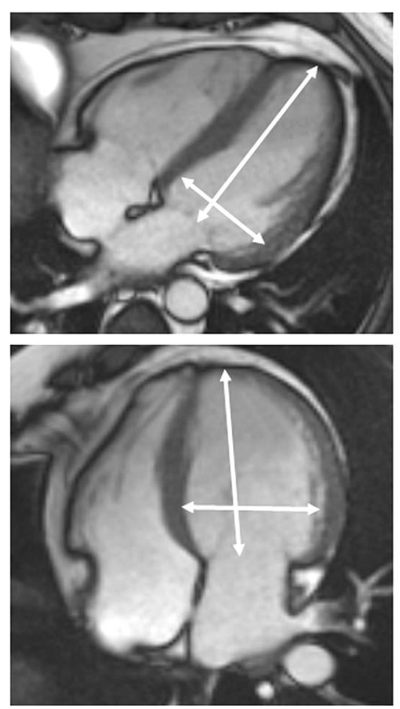
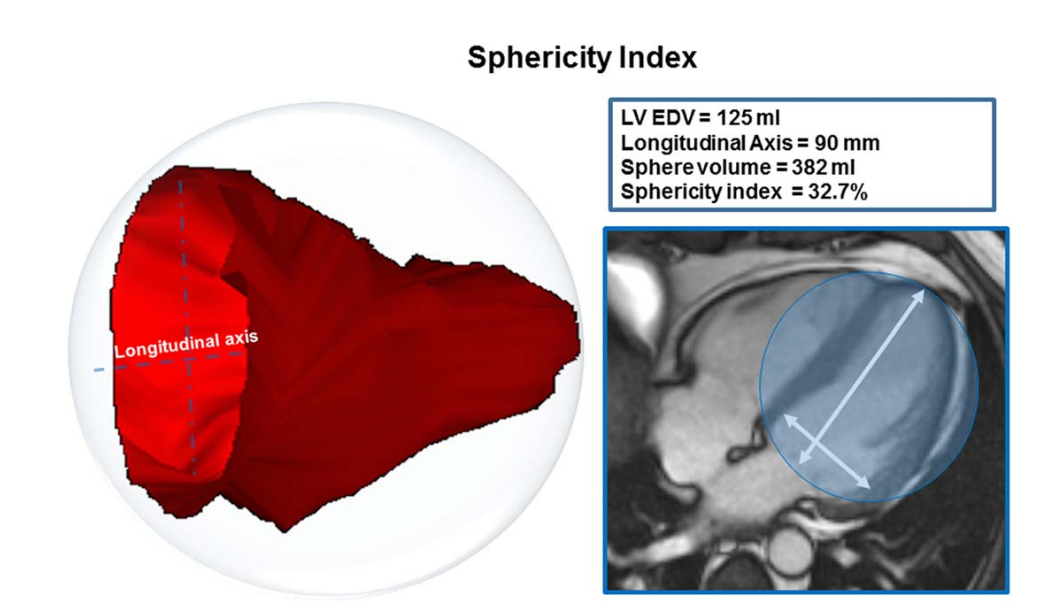
LV diameters and sphericity
For LV diameters, the implementation runs the short-axis diameter helper and the 4-chamber long-axis helper separately at ED and ES. For LV sphericity, it divides LV cavity volume by the volume of a sphere defined by the 4-chamber long-axis diameter:
Copyable formula
SI_LV = V_LV / ((4/3) * pi * (L_4ch / 2)^3)The ED and ES sphericity indices are skipped when the implemented outlier guard detects an extremely high value.
| Geometry route | ED output | ES output | Conditional behavior |
|---|---|---|---|
| Short-axis transverse diameter | LV: D_transverse_ED (sax) [cm] | LV: D_transverse_ES (sax) [cm] | skipped if evaluate_ventricular_length_sax raises ValueError |
| 4ch long-axis diameter | LV: D_longitudinal_ED (4ch) [cm] | LV: D_longitudinal_ES (4ch) [cm] | skipped if evaluate_ventricular_length_lax raises ValueError |
| 4ch transverse diameter | LV: D_transverse_ED (4ch) [cm] | LV: D_transverse_ES (4ch) [cm] | skipped if evaluate_ventricular_length_lax raises ValueError |
| Sphericity index | LV: Sphericity_Index_ED | LV: Sphericity_Index_ES | skipped if index is greater than the implemented outlier guard |
Output columns and units
CardiacNexus writes ventricular structural features into ventricular_volume.csv.
| Display family | Exact output column | Unit | Status | Schema note |
|---|---|---|---|---|
| LV volume | LV: V_ED [mL] | mL | current | ED frame is 0 |
| RV volume | RV: V_ED [mL] | mL | current | ED frame is 0 |
| LV volume | LV: V_ES [mL] | mL | current | ES is LV minimum-volume frame |
| RV volume | RV: V_ES [mL] | mL | current | Uses LV-selected ES frame |
| LV indexed volume | LV: V_ED/BSA [mL/m^2] | mL/m² | current when BSA exists | BSA-dependent |
| RV indexed volume | RV: V_ED/BSA [mL/m^2] | mL/m² | current when BSA exists | BSA-dependent |
| LV indexed volume | LV: V_ES/BSA [mL/m^2] | mL/m² | current when BSA exists | BSA-dependent |
| RV indexed volume | RV: V_ES/BSA [mL/m^2] | mL/m² | current when BSA exists | BSA-dependent |
| LV diameter | LV: D_longitudinal_ED (4ch) [cm] | cm | current when geometry succeeds | 4ch long-axis support |
| LV diameter | LV: D_transverse_ED (sax) [cm] | cm | current when geometry succeeds | short-axis transverse diameter |
| LV diameter | LV: D_transverse_ED (4ch) [cm] | cm | current when geometry succeeds | 4ch transverse diameter |
| LV diameter | LV: D_transverse_ES (sax) [cm] | cm | current when geometry succeeds | short-axis transverse diameter at LV-selected ES |
| LV diameter | LV: D_longitudinal_ES (4ch) [cm] | cm | current when geometry succeeds | 4ch long-axis support at LV-selected ES |
| LV diameter | LV: D_transverse_ES (4ch) [cm] | cm | current when geometry succeeds | 4ch transverse diameter at LV-selected ES |
| LV shape | LV: Sphericity_Index_ED | unitless | conditional current output | Outlier guard may skip |
| LV shape | LV: Sphericity_Index_ES | unitless | conditional current output | Outlier guard may skip |
Output reconciliation
| Evidence layer | Result |
|---|---|
| Implementation source | 16 ventricular-structure outputs documented here: 4 absolute volumes, 4 indexed volumes, 6 LV diameter rows, and 2 LV sphericity rows |
| Output inventory | docs/data/output_column_inventory.yml includes the same 16 ventricular-structure outputs under ventricular_volume.structural_columns |
| Phenotype dictionary | docs/data/phenotype_dictionary.yml links the same 16 ventricular-structure outputs to this page |
| Page output table | all 16 output labels are listed above |
Required upstream inputs
sa.nii.gz,seg_sa.nii.gz,la_4ch.nii.gz, andseg4_la_4ch.nii.gz.- Short-axis segmentation QC passing
sa_pass_quality_control. - BSA lookup for indexed outputs.
Reference ranges with cohort and method context
| Feature | Source | Cohort | Reference value | Status | Note |
|---|---|---|---|---|---|
| LV/RV EDV and ESV | Petersen et al. UK Biobank CMR reference ranges [1] | UK Biobank CMR reference cohort | sex- and age-stratified source rows | Verified context source | Use with segmentation-method context; not copied here as CardiacNexus-specific thresholds |
| CMR chamber volumes | Kawel-Boehm et al. 2020 update [2] | multi-study CMR reference context | study-specific | Verified context source | Papillary/trabecular conventions differ by study |
| LV dimensions and shape context | Aquaro et al. multicenter CMR reference paper [3] | multicenter, multivendor CMR reference cohort | volume, dimension, and functional-parameter context | Verified context source | Current sphericity implementation is geometry-sensitive and uses CardiacNexus 4ch diameter helpers |
Source-located registry status: reference_range_sources.yml maps ventricular EDV/ESV and indexed volume context to Petersen 2017 LV/RV volume reference tables, Kawel-Boehm 2020 CMR reference-update tables, and Aquaro 2017 ventricular dimension/shape context. Exact numeric rows are not copied here as CardiacNexus-specific thresholds because papillary/trabecular conventions, automated segmentation, and indexing choices must be adjudicated before public threshold display.
Disease interpretation
Larger ventricular volumes are common research markers of chamber remodeling in heart failure and cardiomyopathy [1] [2]. Post-infarction remodeling can affect ESV, EDV, and LV shape [3]. These statements are literature context; CardiacNexus does not diagnose disease from a ventricular volume row.
QC caveats and maturity boundary
Basal slice selection, short-axis coverage, label leakage, and ED/ES frame selection can materially change volume outputs. RV values use the LV-selected ES frame, which is common for paired output tables but should be documented when interpreting RV systolic size.
Implementation provenance
Current outputs are implemented in src/feature_extraction/Short_Axis_20209/eval_ventricular_volume.py.
| Feature family | Formula or computational route | Exact output columns | Source code file and function | Upstream dependencies | Conditional behavior | QC artifacts | Schema debt |
|---|---|---|---|---|---|---|---|
| LV/RV absolute volumes | Count seg_sa labels at T_ED = 0 and LV-selected T_ES, then multiply by voxel volume | LV: V_ED [mL], RV: V_ED [mL], LV: V_ES [mL], RV: V_ES [mL] | eval_ventricular_volume.py; ventricular volume loop | sa.nii.gz, seg_sa.nii.gz, spacing metadata | subject skipped if short-axis segmentation fails QC | visualization/ventricle/seg_sa_ED.png, seg_sa_ES.png, raw volume time-series plot | RV ES volume uses LV-selected ES frame |
| BSA-indexed volumes | Divide absolute ED/ES volumes by query_BSA(subject) | LV: V_ED/BSA [mL/m^2], RV: V_ED/BSA [mL/m^2], LV: V_ES/BSA [mL/m^2], RV: V_ES/BSA [mL/m^2] | eval_ventricular_volume.py; indexed volume block | BSA lookup table | subject skipped if BSA is unavailable | output CSV only; inherited volume QC | BSA dependency is external to image files |
| LV diameter geometry | evaluate_ventricular_length_sax and evaluate_ventricular_length_lax at ED and ES | LV: D_longitudinal_ED (4ch) [cm], LV: D_transverse_ED (sax) [cm], LV: D_transverse_ED (4ch) [cm], LV: D_transverse_ES (sax) [cm], LV: D_longitudinal_ES (4ch) [cm], LV: D_transverse_ES (4ch) [cm] | eval_ventricular_volume.py; diameter blocks; utils.cardiac_utils helpers | seg_sa.nii.gz, seg4_la_4ch.nii.gz, image affine axes | diameter rows skipped if helper raises ValueError | sa_ED_diameter.png, la_ED_diameter.png, sa_ES_diameter.png, la_ES_diameter.png; landmark VTK files | current labels encode view and phase but not helper version |
| LV sphericity | LV volume divided by sphere volume from 4ch long-axis diameter | LV: Sphericity_Index_ED, LV: Sphericity_Index_ES | eval_ventricular_volume.py; sphericity blocks | successful 4ch long-axis diameter and LV volume | sphericity row skipped if outlier guard exceeds threshold | same diameter images and landmarks | unitless shape index is geometry-sensitive |
Source audit
- Volume, ED/ES, BSA-indexing, diameter, and sphericity claims were checked against
eval_ventricular_volume.py. - The prior ES-diameter omission was repaired:
LV: D_transverse_ES (sax) [cm],LV: D_longitudinal_ES (4ch) [cm], andLV: D_transverse_ES (4ch) [cm]are now present in the page, output inventory, and phenotype dictionary. docs/data/reference_sources.ymlis present and used as the curated reference-source registry for this page.- One LV sphericity schematic is displayed from a page-local public path and registered in
docs/data/figure_provenance.yml; permission and exact source-panel review remain pending for draft use.
Related pages
References
- Petersen SE, Aung N, Sanghvi MM, Zemrak F, Fung K, Paiva JM, Francis JM, Khanji MY, Lukaschuk E, Lee AM, Carapella V, Kim YJ, Leeson P, Piechnik SK, Neubauer S. Reference ranges for cardiac structure and function using cardiovascular magnetic resonance in Caucasians from the UK Biobank population cohort. Journal of Cardiovascular Magnetic Resonance. 2017;19(1):18. doi:10.1186/s12968-017-0327-9. PMID:28178995; PMCID:PMC5304550.
- Kawel-Boehm N, Hetzel SJ, Ambale-Venkatesh B, Captur G, Francois CJ, Jerosch-Herold M, Salerno M, Teague SD, Valsangiacomo-Buechel ER, Van Der Geest RJ, Bluemke DA. Reference ranges for cardiovascular magnetic resonance in adults and children: 2020 update. Journal of Cardiovascular Magnetic Resonance. 2020;22(1):87.
- Aquaro GD, Camastra G, Monti L, Lombardi M, Pepe A, Castelletti S, Maestrini V, Todiere G, Masci PG, Di Giovine G, Barison A, Dellegrottaglie S, Perazzolo Marra M, Pontone G, Di Bella G. Reference values of cardiac volumes, dimensions, and new functional parameters by MR: a multicenter, multivendor study. Journal of Magnetic Resonance Imaging. 2017;45(4):1055-1067.