Ventricular function
Feature summary
Stroke volume
Difference between end-diastolic and end-systolic cavity volume for LV and RV.
Heart failure (I50)Cardiomyopathy (I42)Disease badges are literature-context navigation only; not diagnoses, CardiacNexus classifiers, or validated phenotype-to-ICD associations.Ejection fraction
Stroke volume normalized to end-diastolic volume and reported as a percentage.
Heart failure with reduced ejection fraction (I50.2)Acute myocardial infarction (I21)Disease badges are literature-context navigation only; not diagnoses, CardiacNexus classifiers, or validated phenotype-to-ICD associations.Cardiac output and index
Stroke volume multiplied by heart rate, with BSA-normalized cardiac index when BSA is available.
Family: functionalUnit family: L/min, L/min/m²Source: Cine short-axis CMRPrimary output group: Flow-rate summariesHeart failure (I50)Disease badges are literature-context navigation only; not diagnoses, CardiacNexus classifiers, or validated phenotype-to-ICD associations.
Ventricular function phenotypes summarize LV and RV pump performance from the same short-axis volume curves documented on Ventricular structure.
- Modality
- Cine short-axis CMR
- UKB source
- Data Field 20209
- Pipeline step
- Short-axis ventricular volume curve extraction
- Outputs
- ventricular_volume.csv, timeseries/ventricle.npz, ventricular volume and derivative QC plots
- Maturity
- Source-audited phenotype page
Clinical question
Stroke volume, ejection fraction, cardiac output, and cardiac index are the core pump-performance summaries derived from ventricular volume curves. They are widely used in CMR reference-range work and clinical interpretation, but CardiacNexus values remain research outputs that require segmentation and cohort context [1] [2].
Anatomical and physiological definition
Stroke volume is the amount of blood ejected during one contraction. Ejection fraction divides stroke volume by end-diastolic volume. Cardiac output multiplies stroke volume by heart rate; cardiac index normalizes cardiac output by body surface area.
Source acquisition and UKB field
The source acquisition is UK Biobank cine short-axis CMR, Data Field 20209. CardiacNexus uses the same ED/ES frame definitions as the ventricular-structure page: ED frame 0 and ES as the LV minimum-volume frame.
What exactly CardiacNexus measures
The functional rows use the structural volumes from the same script. T_ED is frame 0; T_ES is selected from the LV minimum-volume curve and then reused for RV systolic rows.
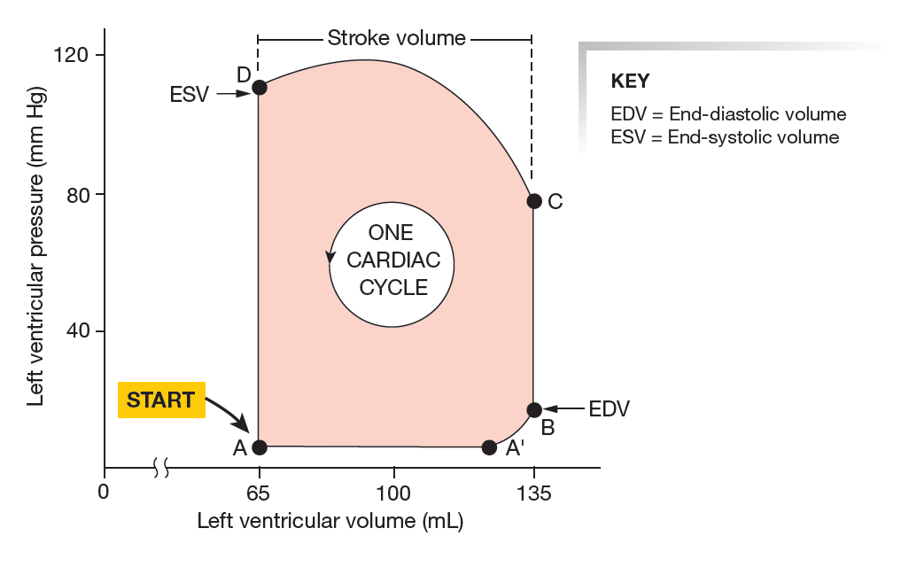
Stroke volume
Copyable formula
SV = V_ED - V_ESEjection fraction
Copyable formula
EF = SV / V_ED * 100Cardiac output and index
CardiacNexus computes heart rate from the number of cine frames and temporal resolution, then multiplies stroke volume by heart rate:
Copyable formula
CO_L_per_min = SV_mL * HR_bpm * 1e-3Copyable formula
CI = CO / BSA| Variable | Definition | Unit | Output dependency |
|---|---|---|---|
LV: V_ED [mL], RV: V_ED [mL] | ED blood-pool volumes from frame 0 | mL | SV and EF denominators |
LV: V_ES [mL], RV: V_ES [mL] | ES blood-pool volumes at LV-selected T_ES | mL | SV numerators |
heart_rate | derived from cine frame count and temporal resolution | beats/min | CO rows |
BSA_subject | returned by query_BSA(subject) | m² | CI rows |
Output columns and units
| Display family | Exact output column | Unit | Status | Schema note |
|---|---|---|---|---|
| LV stroke volume | LV: SV [mL] | mL | current | EDV minus ESV |
| RV stroke volume | RV: SV [mL] | mL | current | EDV minus ESV using LV-selected ES frame |
| LV ejection fraction | LV: EF [%] | % | current | SV divided by LV EDV |
| RV ejection fraction | RV: EF [%] | % | current | SV divided by RV EDV |
| LV cardiac output | LV: CO [L/min] | L/min | current | HR derived from cine timing |
| RV cardiac output | RV: CO [L/min] | L/min | current | HR derived from cine timing |
| LV cardiac index | LV: CI [L/min/m^2] | L/min/m² | current when BSA exists | BSA-dependent |
| RV cardiac index | RV: CI [L/min/m^2] | L/min/m² | current when BSA exists | BSA-dependent |
Output reconciliation
| Evidence layer | Result |
|---|---|
| Implementation source | 8 ventricular-function outputs documented here: LV/RV SV, EF, CO, and CI |
| Output inventory | docs/data/output_column_inventory.yml includes the same 8 outputs under ventricular_volume.functional_columns |
| Phenotype dictionary | docs/data/phenotype_dictionary.yml links the same 8 outputs to this page |
| Page output table | all 8 output labels are listed above |
Required upstream inputs
sa.nii.gzandseg_sa.nii.gz.- Short-axis segmentation QC passing
sa_pass_quality_control. - BSA lookup for cardiac index.
Reference ranges with cohort and method context
| Feature | Source | Cohort | Reference value | Status | Note |
|---|---|---|---|---|---|
| LV/RV SV and EF | Petersen et al. UK Biobank CMR reference ranges [1] | UK Biobank CMR reference cohort | sex- and age-stratified source rows | Verified context source | Use body-size and segmentation-method context; not copied as CardiacNexus-specific thresholds |
| SV, EF, CO, CI | Kawel-Boehm et al. 2020 CMR reference update [2] | multi-study CMR reference context | study-specific | Verified context source | Method conventions vary across studies |
Source-located registry status: reference_range_sources.yml maps SV/EF rows to Petersen 2017 LV/RV stroke-volume and ejection-fraction reference tables and Kawel-Boehm 2020 CMR reference-update context. Cardiac output and cardiac index rows are retained as method-dependent because heart rate, BSA, and automated volume-curve conventions must match before a numeric normal range is promoted.
Disease interpretation
Reduced LVEF is central to many heart-failure and post-infarction discussions, and RV EF/SV are important in pulmonary vascular and right-heart disease contexts [1] [2]. These statements are interpretation context only; CardiacNexus does not assign HFrEF, infarction, or cardiomyopathy diagnoses.
QC caveats and maturity boundary
All ventricular function outputs inherit segmentation and ED/ES frame-selection errors from the ventricular-structure route. Cardiac output and cardiac index also depend on correct temporal metadata and BSA. Preserved EF does not imply normal strain, torsion, wall thickening, or tissue phenotype.
Implementation provenance
Current outputs are implemented in src/feature_extraction/Short_Axis_20209/eval_ventricular_volume.py.
| Feature family | Formula or computational route | Exact output columns | Source code file and function | Upstream dependencies | Conditional behavior | QC artifacts | Schema debt |
|---|---|---|---|---|---|---|---|
| Stroke volume | EDV minus ESV for LV and RV | LV: SV [mL], RV: SV [mL] | eval_ventricular_volume.py; stroke-volume block | LV/RV EDV and ESV from seg_sa.nii.gz | subject skipped if short-axis QC fails before feature calculation | raw and smoothed ventricle_volume*.png; ventricle.npz | RV SV uses LV-selected ES frame |
| Ejection fraction | SV divided by EDV and multiplied by 100 | LV: EF [%], RV: EF [%] | eval_ventricular_volume.py; EF block | SV and EDV rows | inherits volume QC and division dependencies | volume time-series QC | RV EF uses LV-selected ES frame |
| Cardiac output | SV multiplied by derived heart rate and converted to L/min | LV: CO [L/min], RV: CO [L/min] | eval_ventricular_volume.py; CO block | cine frame count, temporal resolution, SV | sensitive to temporal metadata | volume time-series QC | HR is derived from image metadata, not an external ECG value |
| Cardiac index | CO divided by BSA | LV: CI [L/min/m^2], RV: CI [L/min/m^2] | eval_ventricular_volume.py; indexed feature block; query_BSA | BSA lookup table | subject skipped if BSA is unavailable | output CSV only; inherits CO QC | BSA dependency is external to image files |
Source audit
- SV, EF, CO, and CI formula claims were checked against
eval_ventricular_volume.py. - Full ventricular-function output coverage was reconciled across the implementation contract, page text,
docs/data/output_column_inventory.yml, anddocs/data/phenotype_dictionary.yml. docs/data/reference_sources.ymlis present and used as the curated reference-source registry for this page.- One pressure-volume loop schematic is displayed from a page-local public path and registered in
docs/data/figure_provenance.yml; it is broad physiology context only, while current CardiacNexus outputs remain cine-volume-derived.
Related pages
- Ventricular structure
- Ventricular mechanics and strain
- Cine short-axis imaging
- Indexing and normalization
References
- Petersen SE, Aung N, Sanghvi MM, Zemrak F, Fung K, Paiva JM, Francis JM, Khanji MY, Lukaschuk E, Lee AM, Carapella V, Kim YJ, Leeson P, Piechnik SK, Neubauer S. Reference ranges for cardiac structure and function using cardiovascular magnetic resonance in Caucasians from the UK Biobank population cohort. Journal of Cardiovascular Magnetic Resonance. 2017;19(1):18. doi:10.1186/s12968-017-0327-9. PMID:28178995; PMCID:PMC5304550.
- Kawel-Boehm N, Hetzel SJ, Ambale-Venkatesh B, Captur G, Francois CJ, Jerosch-Herold M, Salerno M, Teague SD, Valsangiacomo-Buechel ER, Van Der Geest RJ, Bluemke DA. Reference ranges for cardiovascular magnetic resonance in adults and children: 2020 update. Journal of Cardiovascular Magnetic Resonance. 2020;22(1):87.
- Silverthorn DU, Ober WC, Garrison CW, Silverthorn AC, Johnson BR. Human Physiology: An Integrated Approach. Pearson Education. 2013.